Wandell, B. A., & Smirnakis, S. M. (2009). Plasticity and stability of visual field maps in adult primary visual cortex. Nature Reviews Neuroscience, 10(12), 873–884.
Wang, H. X., & Movshon, J. A. (2016). Properties of pattern and component direction-selective cells in area MT of the macaque. Journal of Neurophysiology, 115(6), 2705–2720.
Wässle, H. (2002). Brian Blundell Boycott, 10 December 1924–22 April 2000. Biographical Memoirs of Fellows of the Royal Society, 48, 53–68.
Wässle, H., Grunert, U., Rohrenbeck, J., & Boycott, B. B. (1989). Cortical magnification factor and the ganglion cell density of the primate retina. Nature, 341(6243), 643–646.
Wässle, H., Puller, C., Muller, F., & Haverkamp, S. (2009). Cone contacts, mosaics, and territories of bipolar cells in the mouse retina. Journal of Neuroscience, 29(1), 106–117.
Watanabe, T., Nбсez, J. E., & Sasaki, Y. (2001). Perceptual learning without perception. Nature, 413(6858), 844–848.
Wathey, J. C., & Pettigrew, J. D. (1989). Quantitative analysis of the retinal ganglion cell layer and optic nerve of the barn owl Tyto alba. Brain, Behavior and Evolution, 33(5), 279–292.
Werner, J. S., & Chalupa, L. M. (Eds.). (2014). The new visual neurosciences. Cambridge, MA: The MIT Press.
Wiesel, T. N. (1982). Postnatal development of the visual cortex and the influence of environment. Nature, 299(5884), 583–591.
Wong, R. O., Meister, M., & Shatz, C. J. (1993). Transient period of correlated bursting activity during development of the mammalian retina. Neuron, 11(5), 923–938.
Wu, K. J. (2018, December 10). Google’s new A.I. is a master of games, but how does it compare to the human mind? Smithsonian.com. Retrieved from www.smithsonianmag.com/innovation/google-ai-deepminds-alphazero-games-chess-and-go-180970981.
Yamins, D. L., & DiCarlo, J. J. (2016). Using goal-driven deep learning models to understand sensory cortex. Nature Neuroscience, 19(3), 356–365.
Yamins, D. L. K., Hong, H., Cadieu, C. F., Solomon, E. A., Seibert, D., & DiCarlo, J. J. (2014). Performance-optimized hierarchical models predict neural responses in higher visual cortex. Proceedings of the National Academy of Sciences, 111(23), 8619–8624.
Zeng, H., & Sanes, J. R. (2017). Neuronal cell-type classification: Challenges, opportunities and the path forward. Nature Reviews Neuroscience, 18(9), 530–546.
Zhang, X., Zhao, J., & LeCun, Y. (2015). Character-level convolutional networks for text classification. In Cortes, C., Lawrence, N. D., Lee, D. D., Sugiyama, M., & Garnett, R. (Eds.), Advances in neural information processing systems 28. Red Hook, NY: Curran.
Zimmerman, A., Bai, L., & Ginty, D. D. (2014). The gentle touch receptors of mammalian skin. Science, 346(6212), 950–954.
Zuccolo, R. (2017, April 3). Self-driving cars – Advanced computer vision with opencv, finding lane lines. Retrieved from https://chatbotslife.com/self-driving-cars-advanced-computer-vision-with-opencv-finding-lane-lines-488a411b2c3d.
Источники иллюстраций
Иллюстрации, не указанные ниже, принадлежат автору и Хаобин Вану.
Глава 1. Три лица: фрагмент рекламного плаката New York City Ballet. Предоставлено Полом Кольником.
Глава 4. Биполярные клетки: рисунок Элио Равиолы.
Глава 5. Баскетболист: с сайта Pixabay@pexel.com; автор Кит Джонсон. Нейроны сетчатки: рисунок Ричарда Маслэнда и Ребекки Рокхилл.
Глава 6. Простая клетка: Hubel, D. (1988), Eye, brain, and vision, New York: Scientific American Library. Сложная клетка: Eye, brain, and vision, New York: Scientific American Library.
Глава 7. Мозг макаки: Yang, R. Visual areas of macaque monkey; http://fourier.eng.hmc.edu/e180/lectures/visualcortex/node6.html. Участки распознавания лиц в мозге мартышки: Hung, C. C., Yen, C. C., Ciuchta, J. L., Papoti, D., Bock, N. A., Leopold, D. A., & Silva, A. C. (2015), “Functional mapping of face-selective regions in the extrastriate visual cortex of the marmoset”, Journal of Neuroscience, 21(35), 1160–1172. Схематические лица: Tsao, D. (2014), “Detail from: The macaque face patch system: a window into object representation”, Cold Spring Harbor Symposia on Quantitative Biology, 79, 109–114.
Глава 10. HOG-изображение: https://medium.com/@ageitgey/machine-learning-is-fun-part-4-modern-face-recognition-with-deep-learning-c3cffc121d78. Схема говорящей нейросети: Rosenberg, C. R., and Sejnowski, T. (1987), “Parallel networks that learn to pronounce English text”, Journal of Complex Systems, 1, 145–168. Классическая нейронная сеть: Bengio, Y. (2016), “Machines who learn”, Scientific American, 314, 46–51. Оригинальный рисунок Джена Кристиансена.
Глава 11. Связи в коре мозга: Felleman, D., and Van Essen, D. (1991), “Distributed hierarchical processing in the primate cerebral cortex”, Cerebral Cortex, 1, 1–47.
Глава 14. Проблема связывания: Sinha, P. (2014), “Once blind and now they see”, Scientific American, 309, 49–55. Изображение любезно предоставлено проектом Пракаша.
Об авторе

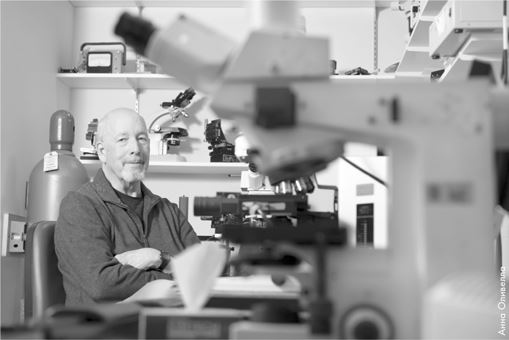
Ричард Маслэнд (1942–2019) был заслуженным профессором офтальмологии кафедры имени Дэвида Гленденинга Когана и профессором нейронаук в Гарвардской медицинской школе. Занимал пост вице-председателя по офтальмологическим исследованиям в Массачусетской клинике болезней глаз, уха, горла и носа при Гарвардском университете, являющейся крупнейшим в мире центром изучения зрения. Более 20 лет преподавал в Гарвардской медицинской школе курс нейробиологии, который дважды признавался лучшей учебной программой. Был членом Американской ассоциации содействия развитию науки (AAAS), исследователем в Медицинском институте Говарда Хьюза, лауреатом премии Проктора за научные достижения, премии Alcon Research и многих других. Работы Маслэнда открыли новые горизонты в изучении нейронных сетей сетчатки глаза.


